


Radiological diagnostics

The ARTIMEDICA clinic has a thriving Digital Tomography and Pantomography Laboratory, where an electroradiologist ensures correct and reliable RVG diagnostics.
We offer patients at the ARTIMEDICA clinic in Wrocław on-site CT scans, pantomographic images (panoramic X-rays), cephalometric images, and RVG point images of teeth.
Cone beam computed tomography (CBCT)
Tomographic examination is currently the most accurate source of information about internal tissues available in non-invasive diagnostics.
For various types of treatment, we also perform tomographic examinations. CBCT tomographic images are useful in: implant treatment, maxillofacial surgery, dental prosthetics, endodontics, orthodontics, periodontology, functional diagnostics and surgical diagnostics for maxillofacial joint dysfunction.
At the ARTIMEDICA Centre, we perform tomographic imaging of:
the mandible and/or maxilla (in a single image during a single scan),
the maxillary sinuses,
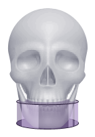
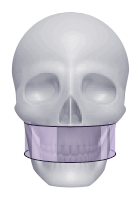
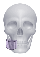
Tomographic examination areas
-

- 11 x 8cm
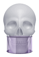
-
- 11 x 5 cm dolne
-
- 11 x 5 cm górne
-
- 5 x 5 cm
Panoramic X-rays of teeth
A pantomographic image allows the dentist to assess the overall condition of the teeth and bones, and thus determine the appropriate treatment plan.
A pantomographic image of the teeth, commonly referred to as a panoramic X-ray, is essential for starting dental and orthodontic treatment. It allows the dentist to assess the overall condition of the entire dentition and bones, and as a result, determine the appropriate treatment plan.
Thanks to the use of the modern Hyperion X9 tomograph, we take pantomographic images in as many as 20 projections, including:
- PAN – standard or paediatric pantomogram;
- DENT – full or partial view of the teeth;
- SIN – frontal or lateral view of the maxillary sinuses;
- TMJ – lateral or lateral-posterior view of the temporomandibular joints, taken at different angles.
A panoramic image provides the dentist with an overview of the condition of the patient’s entire dentition. Detailed information about the health of each tooth is provided by spot images of the teeth, which are also taken at our clinic.
Cephalometric images
Cephalometric radiographs play a very important role in orthodontic treatment, as they provide information about the structure of the patient’s skull and jawbone.
Before starting orthodontic treatment, the dentist needs to examine not only a comprehensive image of the patient’s teeth (panoramic X-ray), but also the structure of the patient’s skull and jawbone. This information is provided by a cephalometric X-ray, i.e. a lateral radiographic image of the teeth and the facial part of the skull.
Thanks to the use of high-class technology, we take cephalometric images in 7 projections, including:
- a cephalogram in a standard or paediatric lateral-posterior view;
- cephalogram in posterior-anterior or anterior-posterior view;
- X-ray image of the hand (wrist).
RVG images of teeth (spot images)
Spot X-rays of teeth allow the condition of the root, neck and crown of the tooth to be assessed, which is crucial in dental treatment.